Lumencor: MIRA on the brain
At Virginia Tech’s School of Neuroscience, Professor Ian Kimbrough provides students with hands-on experience of real-world neuroscience research techniques including immunohistochemistry, neuroanatomy, and microsurgery, just to name a few.
Fluorescence microscopy is the fundamental and essential tool underpinning these techniques. Professor Kimbrough’s teaching laboratory has a fleet of ~20 Nikon Eclipse E200 fluorescence microscopes equipped with Lumencor MIRA light engines®.
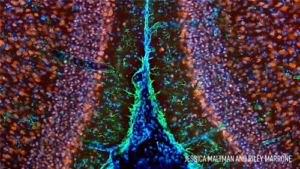
The MIRA light engine provides powerful, stable, and responsive fluorescence excitation for visualization of coronal mouse slices to elucidate cellular definition of mouse brain anatomy. A video presentation including more images can be viewed here. The coronal mouse brain sections were stained using immunohistochemical techniques to fluorescently label various brain cell types and structures: neuronal nuclei (blue), neurons (red), astrocytes (green), and NISSL bodies (yellow).